Treatment of cartilage injuries depends on numerous factors such as symptoms, age of the patient, and the size, depth and location of the cartilage ulcer. Selecting the right treatment option is a complex process, best carried out in consultation with an orthopaedic surgeon. Some of the options are listed below:
Debridement
Simple, small and shallow cartilage ulcer may only require debridement or "cleaning up" to smooth out the rough surface or any shallow unstable flaps of cartilage. This can be performed at the time of arthroscopy.
Microfracture
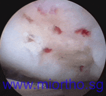
This is also performed at the time of arthroscopy. Small holes are made in the base of the ulcer. This causes bleeding from the bone marrow which allows for healing. The ulcer usually fills in with a type of scar tissue called fibrocartilage, which is different from normal joint cartilage (hyaline cartilage). Success rates are generally still quite high, particularly for smaller ulcers of an area of less than 1-2 cm squared.
The picture on the right demonstrates microfracture of a cartilage ulcer, with bleeding from the small holes made in the base of the ulcer.

Cartilage resurfacing
Cartilage resurfacing involves attempting to replace the ulcer with cartilage that is the same as, or similar to normal joint cartilage (hyaline cartilage). One option is mosaicplasty, which uses small circular plugs of cartilage and bone taken either from the patient, or from cadaveric tissue. These plugs are then applied to the ulcer like mosaic tiles - hence the name, mosaicplasty.

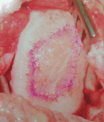
Another option is using the patient's own cells, either cartilage cells or bone marrow stem cells, to attempt to re-grow new cartilage in the ulcer. This technique is called Autologous Chondrocyte Implantation (ACI). Typically this requires 2 separate operations, the first to harvest the cells, and the second to implant them. Culturing the cells usually takes between 3 to 6 weeks. The picture to the right shows ACI performed on a 3 cm square cartilage defect.
The implantation surgery is usually open traditional surgery, in which the cells are impregnated in a collagen scaffolding (that looks like a piece of wet tissue paper), and this is pasted into the cartilage defect.
